
menu
cancel
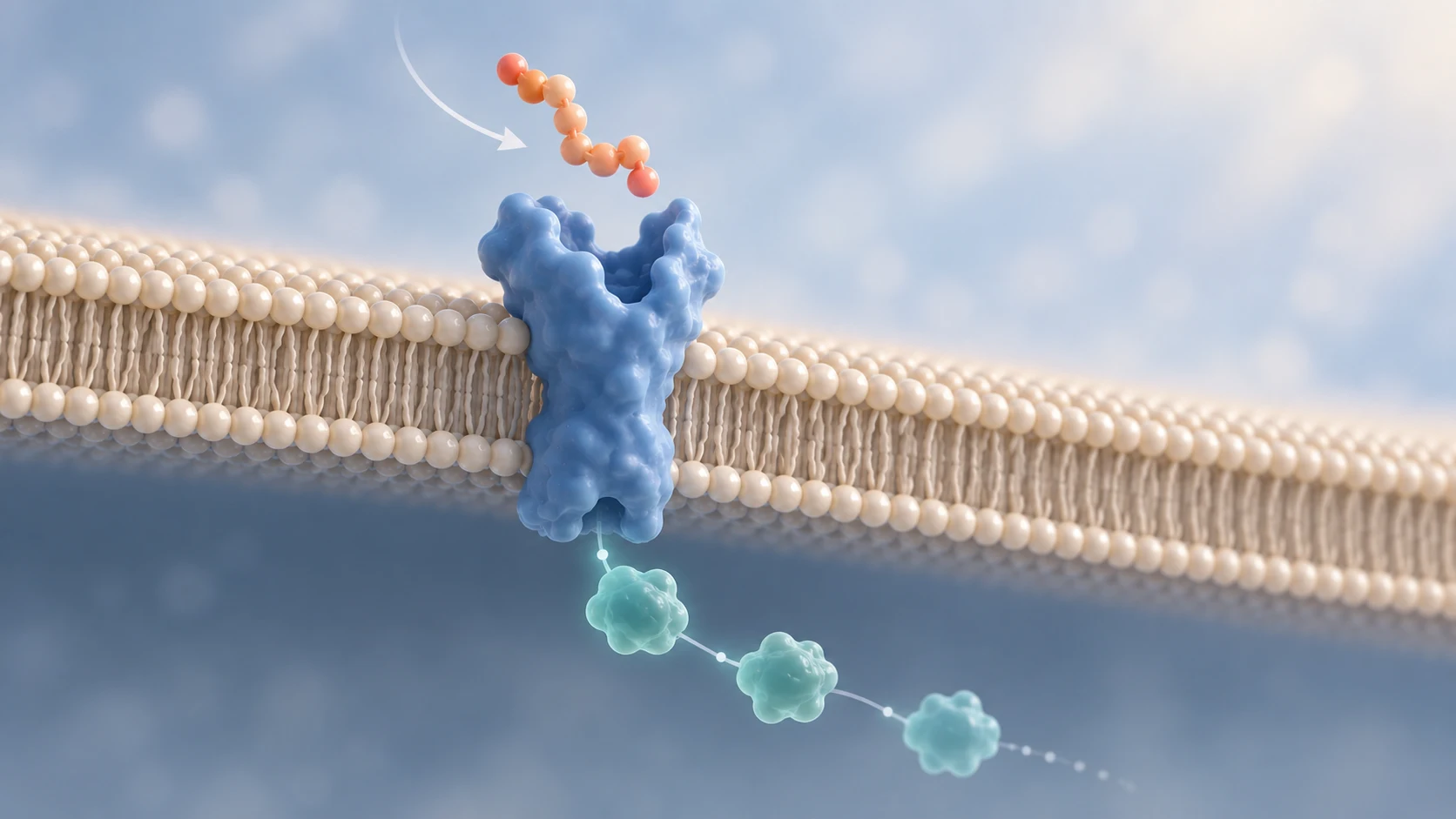
Peptides do not force every cell to perform the same task.
They carry signals.
A cell can respond only when several conditions line up. The peptide has to reach the cell in an active form. The cell has to display a compatible receptor. That receptor has to convert binding into an intracellular signal. The machinery downstream then has to process that signal in the context of the cell's current state.
That sequence is the foundation of peptide biology. It is also why the phrase 'this peptide activates this pathway' rarely tells the whole story.
Receptors are dynamic proteins, not simple on-and-off buttons. Signaling pathways branch, amplify, overlap, adapt, and shut down. The same peptide may produce different responses in different tissues because the biological context is different.
Here is a clear model for how peptide communication works, where the complexity enters, and why a mechanism should never be confused with a guaranteed outcome.
A peptide is a chain of amino acids. Some peptides act as biological messengers, including hormones, neuropeptides, growth-related signals, and local signaling molecules.
The peptide itself does not contain a written command. Its amino acid sequence creates a particular chemical surface, shape, charge pattern, and flexibility. Those features influence which molecules the peptide can interact with and how strongly those interactions occur.
In signaling biology, the peptide is often called a ligand. A ligand is a molecule that binds to another molecule, commonly a receptor. Binding can stabilize a receptor in an active, partially active, inactive, or otherwise distinct conformation depending on the receptor and ligand involved.
That distinction matters. Contact is not the endpoint. The important question is what the receptor and the cell do after contact occurs.
A receptor is a protein built to recognize certain chemical signals and translate them into biological activity. Many peptide signals act through receptors located on the cell surface because their size and chemical properties limit passive movement through the lipid membrane.
The lock-and-key analogy is useful, but incomplete. A receptor is not a rigid lock and a peptide is not a rigid key. Both molecules can move. Binding changes the balance among receptor shapes, and those shapes can favor different signaling partners or pathways.
Receptor expression also creates biological selectivity. A cell that does not express the relevant receptor will not interpret the signal through that receptor. A cell with abundant receptor, a different receptor subtype, or a different set of accessory proteins may respond differently.
This is one reason a peptide name alone cannot predict an effect across the whole body. The distribution of receptors helps define where a signal can be heard. The cellular machinery helps define how it is interpreted.
A useful way to understand peptide signaling is to separate it into five connected stages.
Peptides do not all use one receptor family. The receptor's architecture determines how an outside binding event is connected to the inside of the cell.
Many peptide hormones signal through G protein-coupled receptors, or GPCRs. These receptors cross the cell membrane seven times. When a peptide stabilizes an active receptor conformation, the receptor can engage G proteins and other signaling partners.
Depending on the receptor and cell, this may change cyclic AMP, calcium, lipid-derived messengers, protein kinase activity, ion channels, or gene regulation. Class B GPCRs include receptors for peptide hormones such as glucagon, calcitonin, and parathyroid hormone.
The phrase 'binds a GPCR' therefore does not identify one universal response. It identifies the beginning of a family of possible signaling events.
Other peptide signals act through receptor tyrosine kinases. These receptors have an extracellular binding region, a membrane-spanning segment, and an intracellular enzyme domain.
Ligand binding changes receptor organization and kinase activity. The receptor then phosphorylates specific amino acid residues, creating docking sites for proteins that assemble branching signaling networks. PI3K-AKT, RAS-MAPK, and phospholipase C pathways are common examples, but their use depends on the receptor and cellular system.
Peptide and protein signals can also act through cytokine receptors, receptor guanylyl cyclases, serine/threonine kinase receptors, and receptor systems that depend on coreceptors or accessory proteins. Some peptides interact with more than one receptor subtype.
The receptor family is not a technical footnote. It shapes the speed, duration, location, and type of biological response that researchers should expect to measure.
Once a receptor is activated, the message is relayed through interacting proteins and small intracellular messengers. Common mechanisms include phosphorylation, changes in cyclic AMP, changes in calcium concentration, lipid signaling, protein trafficking, and altered gene transcription.
These cascades can amplify a signal. One activated receptor may influence multiple downstream molecules, and each of those molecules may influence others. Amplification helps cells respond to small changes in an external signal.
The same network also contains restraints. Phosphatases remove phosphate groups. Inhibitory proteins limit pathway activity. Feedback loops can strengthen, reshape, or reduce the response. Signals from other receptors may converge on the same intracellular nodes.
This is why a pathway diagram should be read as a map of possible relationships, not as proof that every arrow is equally active in every cell.
A biological signal is interpreted by a system, not by the peptide alone.
Two cell types may express different amounts of the same receptor. They may use different G proteins, kinases, adaptor proteins, phosphatases, transcription factors, or metabolic enzymes. Their energy state, developmental stage, exposure history, and surrounding signals may also differ.
As a result, the same receptor can connect to different downstream priorities. One cell may change secretion. Another may change nutrient handling. Another may alter gene expression, movement, survival signaling, or growth.
The response can also change with time. A brief signal may alter enzyme activity within seconds. A sustained signal may change receptor availability or gene expression over minutes to hours. Repeated exposure may produce adaptation or desensitization.
Context is not noise around the mechanism. Context is part of the mechanism.
Insulin provides a familiar example of peptide-to-receptor communication without reducing the biology to one arrow.
Insulin binds the insulin receptor, a receptor tyrosine kinase. Structural studies show that insulin binding reorganizes the receptor and stabilizes an active arrangement. The intracellular kinase domains then phosphorylate the receptor and recruit signaling substrates.
Two major signaling branches are often described. One involves insulin receptor substrate proteins, PI3K, and AKT and is central to many metabolic effects. Another involves adaptor proteins that connect the receptor to RAS-MAPK signaling and other growth-related processes.
In skeletal muscle and adipose tissue, insulin signaling can promote movement of GLUT4-containing vesicles toward the cell membrane, increasing the cell's capacity for glucose uptake. In the liver, insulin participates in a different tissue-level program involving glucose production, glycogen metabolism, lipid metabolism, and indirect signals from other tissues.
Insulin is one peptide. Its coordinated biological effect emerges from receptor activation across several tissues, different intracellular machinery, feedback, and communication between organs.
That is the larger lesson. A receptor explains where a signal begins. It does not, by itself, explain the whole organism.
Peptide discussions often collapse several different measurements into the word 'works.' That shortcut creates confusion.
A ligand may bind tightly but activate weakly. A partial agonist may produce a lower maximum response than a full agonist in the same test system. Two ligands can act at the same receptor yet favor different signaling profiles, a concept often described as functional selectivity or biased agonism.
None of these terms should be treated as a universal quality ranking. Their meaning depends on the assay, receptor system, endpoint, timing, and biological context.
Biology depends on regulation. Receptors and pathways evolved to operate within ranges, respond to timing, and interact with feedback systems.
A stronger or longer signal may increase one measured response, but it may also recruit additional pathways, increase off-target activity, trigger counter-regulation, or cause the cell to reduce its sensitivity. The preferred response is therefore not always the largest laboratory signal.
The right question is not simply whether a peptide activates a receptor. Researchers need to ask which receptor states are stabilized, which pathways are engaged, at what concentrations, for how long, in which cells, and with what downstream consequences.
Cell signaling needs an off-switch as much as an on-switch.
The peptide may be degraded or cleared. The receptor may be phosphorylated, uncoupled from its signaling partner, pulled into the cell, recycled, or directed toward degradation. Intracellular messengers may be broken down, and phosphatases can reverse phosphorylation events.
For many GPCRs, receptor kinases and arrestin proteins help reduce continued G protein signaling after activation. Internalization may reduce surface receptor availability, while arrestins can also participate in their own signaling and trafficking functions.
These processes help explain why duration of exposure, timing between signals, and receptor history can change what researchers observe. A single snapshot may miss the adaptation that follows.
Mechanistic evidence is valuable. It can show that a peptide binds a target, changes receptor activity, or influences a pathway in a defined experimental system.
But each level of evidence answers a different question. A purified receptor study can reveal molecular interaction. A cell experiment can show pathway activity in that cell model. An animal study can add metabolism, tissue distribution, and whole-organism biology. Human clinical research is needed to evaluate safety and effectiveness in people for a defined use.
Translation can fail because exposure is insufficient, the relevant receptor distribution differs, compensatory pathways intervene, the model does not represent the human condition, or the measured laboratory endpoint does not predict a meaningful clinical result.
A mechanism makes a hypothesis more specific. It does not make the conclusion inevitable.
Binding shows molecular interaction under the tested conditions. It does not establish tissue exposure, pathway selectivity, dose-response behavior, safety, or a meaningful human outcome.
Affinity is only one property. Activation, selectivity, duration, distribution, and the biological endpoint still matter.
Receptors often connect to branching networks. The same receptor can recruit different signaling partners, and pathways can interact with signals from other receptors.
Cells differ in receptor expression and downstream machinery. A peptide can reach two tissues and still produce different responses.
Endogenous signaling is controlled by concentration, location, timing, processing, binding proteins, clearance, and feedback. Changing those conditions can change the response.
Before accepting a mechanism claim, ask:
1. Which receptor or receptors were directly tested?
2. Was binding measured, receptor activation measured, or only a downstream marker observed?
3. Which cell type, tissue, species, and experimental model were used?
4. Were the peptide concentrations and exposure times biologically realistic?
5. Which signaling pathways and endpoints were measured?
6. Were selectivity, off-target activity, desensitization, or feedback evaluated?
7. Does the evidence come from purified proteins, cells, animals, or human studies?
8. Does the conclusion stay within what that level of evidence can actually establish?
The more confidently someone predicts a human result, the more human evidence you should expect to see.
Peptide communication begins with molecular recognition, but it does not end there.
The peptide must reach the right place. A compatible receptor must be present. Binding must produce a receptor state that can engage intracellular machinery. The signal must then move through a network shaped by cell type, timing, feedback, and competing inputs.
The scientific standard is clear. Receptor binding can support a mechanism, and a pathway can support a hypothesis. Neither replaces evidence of what actually happens in people.
Start with the signal. Follow the receptor. Trace the pathway. Then ask whether the outcome was truly demonstrated.
3. Lemmon MA, Schlessinger J. Cell signaling by receptor tyrosine kinases. Cell. 2010;141(7):1117-1134.
7. U.S. Food and Drug Administration. The Drug Development Process.
This article is for general educational andinformational purposes only. It does not constitute medical, scientific,laboratory, regulatory, diagnostic, or treatment advice.
Peptide behavior depends on the exactmolecule, formulation, route, exposure, receptor system, cell type, tissue,study design, and individual clinical context. Mechanistic findings frompurified proteins, cell models, or animals should not be treated as proof ofsafety or effectiveness in people.
Do not use this article to select, obtain,prepare, dose, combine, administer, or discontinue any product or treatment.Consult qualified medical and scientific professionals and follow applicableproduct labeling and regulatory requirements.